Fungi identification via a Compound Microscope
|
Fungi features can look very beautiful under a compound microscope with a 1000x oil immersion lens. The latest means of photographing microscopic images and viewing them on your computer or smart device offers fungi enjoyment during the winter months.
Different species of fungi can look very similar and another way of confirming a find's identity is to examine its features under a microscope. The easiest and first thing to look at are the spores, which differ in size, shape and ornamentation. You can also look at Asci, clamp connections, basidia and cystidia. To see microscopic fungi parts like spores and ascii clearly you will need a good quality 1000x magnification compound microscope with an oil immersion lens.
A Compound microscope combines the magnification power of a lense that rotates above your slide with that of the ocular lens you look through.
A compound microscope usually has four objective lenses with 4x, 10x,20-40x and 100x magification and a 10x magnification ocular lens making a total magnification of 1000 times.
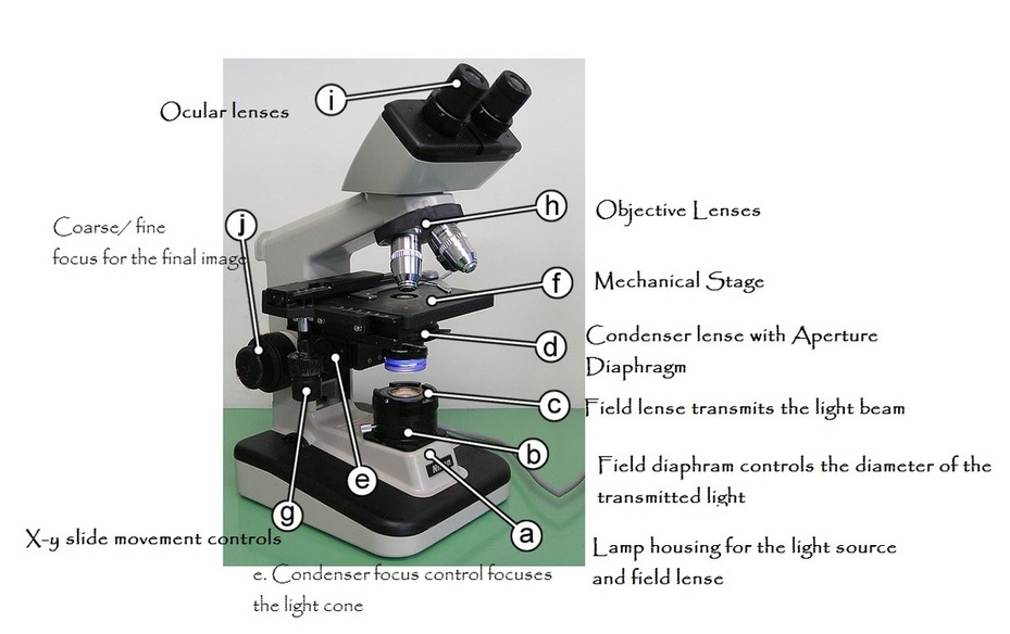
Take a moment to find the ocular (i) and objective (h) lenses on the diagram. |
High Microscope Magnification Needs good Resolution
Quite a few compound microscopes are advertised as having 2000 or more times magnification. However, don't be lured by promises of magnification much greater than 1000 times. It can be easier to measure spores at greater magnification but the images will be blurred and lack detail because increased magnification does not increase resolution. This will be the problem with the new 1000x iphone microscope attachment.
The principle is the same as enlarging a photo without increasing the number of megapixels within it. The photo will pixelate rather than show more detail. Resolution Capacity
A microscope's resolution capacity results from the quality of the available light, of the condensor and objective lenses (c,d,h,l) through which the light passes, and the mechanisms for focusing (j,e) and adjusting the strength, diameter (b ,d) and angle (d) of that light.
Modern compound microscopes supply light through a lamp mounted underneath the glass slide on which your fungi specimen is mounted, at point a on the diagram. Click here for more on the types of lighting you can choose
|