FungiOz - Australian fungi & more
Copyright (2014)
  • Home
    • FungiOz Goals, Policies and contact form
    • 12 apostles visitor
    • Fungi resources
    • Fungi Books
  • Australian fungi
    • Australian Gilled Fungi menu >
      • Australian Amanitas >
        • 3 large Amanitas
        • Amanita cinnereoannnulosa
        • Amanita farinacea
        • Amanita Xanthocephala
      • Australian Web-cap fungi >
        • Cortinarius Archeri
        • Cortinarius Austroalbidus
        • Cortinarius Lavendulensis
        • Dermocybe austroveneta
        • Dermocybe canaria
        • Dermocybe splendida
        • Cortinarius sinapicolor
      • Australian Russulas -Brittle stems
      • Agaricus mushrooms
      • Lactarius Mushrooms
    • Australian crinkled fungi
    • Australian resupinate fungi
    • Australian coral fungi >
      • Australian Clavarias
      • Australian Ramarias
    • Australian Sarcodon
    • Australian Fungi-Boletes >
      • Australian Austroboletes fungi
      • Boletellus obscurecoccineus
    • Australian globular fungi
  • Forays
  • Apps
    • FungiOz App >
      • 3 Step Fungi ID
      • Add to fungi knowledge
      • Get Fungi0z Now
    • Audubon Mushrooms
    • Mushrooms Pro App >
      • Interactive features
      • Review summary
  • Terms
    • Fungi Cap Shapes and Cap Margin shapes >
      • Fungi Cap Margin Types
    • Fungi Stem Shapes >
      • Fungi Stem Surfaces
      • Fungi Stem bases and Ring types
    • Fungi Gill Types and Edges >
      • Fungi Gill to Stem Relationships
    • Fungi Surfaces: Scaly, with Warts or Smooth
    • Fungi Surfaces with hair
  • Shop
    • Carry-all Tote Bags
    • Fungi Coasters
    • Cards
    • Games & Posters
    • Fungi photo Mugs
    • Travel mugs
    • Phone Covers
    • Fungi Designer T-shirts
    • Designer Fungi Aprons for Gourmet Cooks
    • Calendars >
      • DIY Calendar - Fungi
    • Throw Cushions
  • Blog
  • Fungi Imaging
    • Photographing Fungi
    • Iphone microscopes
    • Parts of a Compound Microscope
    • Microscope light sources
    • How to assess a Microscope Condenser lens
    • All about Oil Immersion Lenses
    • About Objective Lenses >
      • Microscopes with Achromatic Objectives
      • Microscopes with Plan objectives

All about Light Sources in
Compound Microscopes

Pros + Cons of tungsten, LED, florescent & halogen globes for microscopic fungi imaging,

Join an Australian Fungi Foray
Types of microscope light sources
Picture
Resolution increases with the quality of the light that passes through  your specimen.   

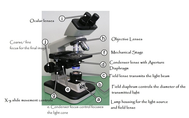
That is why old or cheap microscopes that use a rotating mirror to catch ambient light lead to unsatisfactory images  especially at higher magnifications. 


You will need a built-in, dimmable electric light source, as shown on point A of the above diagram.

Resolution is improved when shorter wavelengths of light are used to illuminate your specimen because shorter wavelengths are more intense.  Different type of light have different wave lengths.

Tungsten globes

A hot light, like a tungsten globe is likely to dry out the specimen. Tungsten globes are cheap but are often hard to replace in older microscope models because they were not standardised when they became popular for built in microscope lighting.

Fluorescent and LED globes  are cool, cheap to run and  long-lasting.


  • Fluorescent globes produce more natural  looking images. 

LED globes

  • Are usually dimmable

  • Are available in various versions of white light with those having more violet light  having the  shortest wavelengths.

  • But LED globes are often wired into their own circuit making them hard to replace.

Halogen globes

  • Medical and research microscopes are likely to have halogen bulbs.

  • Their light is intense but produce natural colours.

  • They usually have dimmer controls. 

  • A  binocular scope is best with halogen or LED lighting  because of the extra brightness needed to light  both tubes.
Other Pointers
  • Check that the microscope globe and fitting is a standard type that preferably can be purchased for common applications.

  • Otherwise  you may not be able to replace the globe when it fails some years down the track.
     
  • Make sure the light is dimmable rather than just having an on and off  switch.

    There should be a moveable diaphragm  called a Field Diaphram, located near  the light source as in the diagram point  b.  

    It will allow you to  control the diameter of the light beam  to eliminate stray light and send all the light upwards toward your specimen. 

Click here to learn about other factors that increase the quality of microscope light and hence the resolution of observed images.

Picture

  Fungi Catalogue menu  
Illustrated mycology terms
       Fungi giftware     
Picture
Browse fungi Design gifts
Home    Apps   Aussie fungi  Forays   Terms    Blog   Shop  Imaging    Privacy Policy   Links   Contact 
Copyright 2014